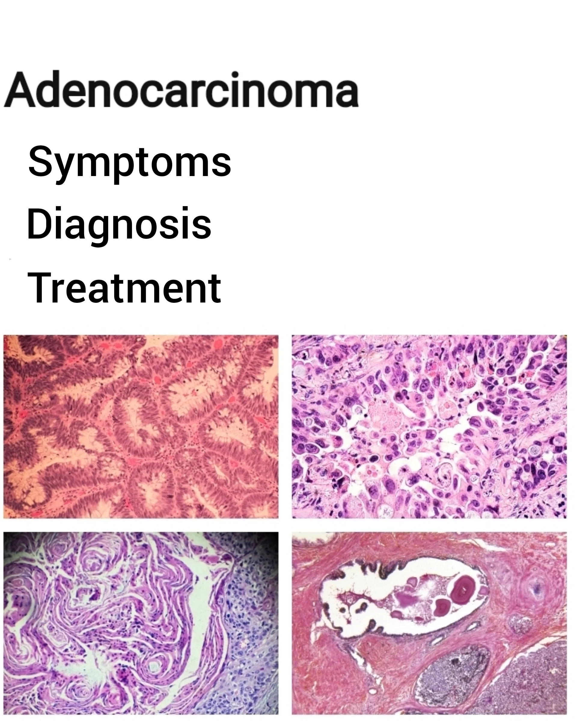
What is Adenocarcinoma?
Lung adenocarcinoma is a form of non-small cell lung cancer. It develops when abnormal lung cells proliferate uncontrollably and create a tumor. Tumor cells can eventually spread (metastasize) to other areas of the body, including the
lymph nodes in and around the lungs
liver
bones
adrenal glands
The most frequent type of lung cancer is adenocarcinoma. It is more frequently encountered among smokers. It is, nonetheless, the most prevalent type of lung cancer in nonsmokers. Additionally, it is the most frequent type of lung cancer in women and adults under the age of 45. Adenocarcinoma, in comparison to other kinds of lung cancer, is more likely to be localized. If the cancer is genuinely confined, it may react better to treatment than other types of lung cancer.
As is the case with other types of lung cancer, your risk of developing adenocarcinoma increases if you smoke.
Smoke. Cigarette smoking is by far the most significant risk factor for lung cancer. Indeed, cigarette smokers are 13 times more likely than nonsmokers to acquire lung cancer. Cigar and pipe smoking are almost as probable as cigarette smoking to cause lung cancer.
Inhale tobacco smoke. Nonsmokers who inhale a cigarette, cigar, or pipe smoke are at an elevated risk of developing lung cancer.
Are at risk of being exposed to radon gas. Radon is a radioactive gas that is colorless and odorless. It is created in the ground. It seeps into the basements of homes and other structures, contaminating drinking water. Exposure to radon is the second most common cause of lung cancer. It is unknown whether elevated radon levels contribute to nonsmoker lung cancer. However, radon exposure does contribute to an elevated risk of lung cancer among smokers and those who inhale large volumes of the gas on a daily basis (miners, for example). A radon testing kit can be used to determine the radon levels in your house.
Are asbestos-exposed. Asbestos is a mineral that is utilized in a variety of products, including insulation, fireproofing materials, floor and ceiling tiles, and vehicle brake linings. Individuals who are exposed to asbestos on the job (miners, construction workers, shipyard employees, and some auto mechanics) have a significantly increased risk of lung cancer. Individuals who live or work in deteriorating buildings that contain asbestos are also at an elevated risk of lung cancer. Along with an increased chance of adenocarcinoma, individuals exposed to asbestos have an increased risk of developing mesothelioma. This is a form of cancer that begins in the lung tissue. Mesothelioma can also develop in the tissue that surrounds abdominal organs.
At work, they are exposed to more cancer-causing chemicals. Uranium, arsenic, vinyl chloride, nickel chromates, coal products, mustard gas, chloromethyl ethers, gasoline, and diesel exhaust are just a few examples.
What are the Symptoms of Adenocarcinoma?
Many persons with lung adenocarcinoma or other kinds of lung cancer exhibit no symptoms. It may be found through a chest x-ray or CT scan for screening purposes or for another medical cause.
All types of lung cancer, including adenocarcinoma, present with comparable symptoms. They include the following:
a persistent cough
coughing up blood or mucous wheezing breathlessness difficulty breathing
ache in the chest fever discomfort when swallowing hoarseness
loss of weight
Appetite deficit.
Other symptoms may occur if the malignancy has progressed beyond the lungs. For instance, you may get bone discomfort if cancer has migrated to your bones.
Diagnosis of Adenocarcinoma
Your physician will begin by taking a medical history. He or she will inquire about your smoking habits and whether you share an apartment with another smoker. Additionally, your doctor will inquire about any possible occupational exposure to asbestos or other cancer-causing substances.
Following that, he or she will conduct imaging tests to screen for masses in your lungs. Typically, a chest x-ray will be performed first.
A CT scan will be performed if the x-ray reveals anything abnormal. The scanner takes numerous images as it goes around you. The photos are then combined by a computer. This produces a more detailed image of the lungs, which enables physicians to confirm the size and location of a mass or tumor.
Additionally, a magnetic resonance imaging (MRI) or positron emission tomography (PET) scan may be performed.
MRI scans produce comprehensive images of the body's organs, but they do so through the use of radio waves and magnets, not x-rays.
PET scans examine the function of the tissue rather than its anatomical structure. On a PET scan, lung cancer and a variety of other malignancies exhibit high metabolic activity. A vein is injected with radioactive sugar. Cancer cells are more active and require more sugar than normal cells. This causes cancerous regions to appear brighter on the scan.
If cancer is detected as a result of these images, additional tests will be performed to confirm the diagnosis, define the type of cancer, and determine if cancer has spread. Among these tests are the following:
Sputum sample – Mucus that has been coughed up is examined for cancer cells.
Biopsy - In a laboratory, a sample of abnormal lung tissue is extracted and analyzed under a microscope. Frequently, the tissue is collected during a bronchoscopy. Surgical exposure of the suspect area, on the other hand, maybe essential.
Bronchoscopy – This procedure involves the passage of a tube-like tool down the throat and into the lungs. A camera attached to the end of the tube enables doctors to look for cancer and biopsy a small bit of tissue.
Mediastinoscopy – A tube-like instrument is used to biopsy lymph nodes or tumors between the lungs during this operation. (This region is referred to as the mediastinum.) A biopsy taken in this manner can be used to establish the type of lung cancer and whether it has spread to the lymph nodes.
A CT scan can be used to identify a questionable location via fine-needle aspiration. After that, a small needle is put into that area of the lung. The needle extracts a small amount of tissue for laboratory evaluation. This allows for the diagnosis of the cancer type.
Thoracentesis – If a build-up of fluid in the chest occurs, it can be drained using a sterile needle. Following that, the fluid is screened for cancer cells.
VATS (video-assisted thoracoscopy) – In this surgery, a surgeon makes an incision in the chest and inserts a flexible tube with a video camera attached to the end into the chest. He or she can then examine the area between the lungs and the chest wall for malignancy. Additionally, abnormal lung tissue can be removed.
CT, PET, and bone scans – These imaging procedures can detect the spread of lung cancer to the brain, bones, or other regions of the body.
Thoracotomy. On occasion, a bigger incision in the chest may be necessary to obtain tissue for laboratory analysis.
After a cancer diagnosis is made, it is given a "stage." The stage identifies the size and extent of the tumor. Stages I through III are further classified as "A" and "B." Tumors in stage I are tiny and have not infiltrated adjacent tissues. Tumors in stages II and III have invaded nearby tissues and/or organs and spread to lymph nodes. Tumors in stage IV have expanded outside the chest.
Duration Estimated
Lung adenocarcinoma will continue to grow and spread unless and until it is treated.
Prevention of Adenocarcinoma
To lower your risk of developing adenocarcinoma or other types of lung cancer,
Avoid smoking. If you are already a smoker, speak with your physician about getting the assistance you need to quit.
Avoid exposure to secondhand smoking. Select restaurants and motels that are smoke-free. Inviting guests to smoke outdoors is a good idea, especially if you have children in your home.
Reduce your radon exposure. Conduct a radon gas test on your home. A radon concentration of more than 4 picocuries/liter is considered dangerous. If you have a private well, you should also have your drinking water tested. Radon test kits are commonly accessible.
Reduce asbestos exposure. Due to the fact that there is no safe amount of asbestos exposure, any exposure is considered excessive. If you live in an older home, check to see if any asbestos-containing insulation or other materials are exposed or degrading. Asbestos must be properly removed or sealed in certain areas. If the removal is not performed correctly, you may be exposed to more asbestos than you would have been had the asbestos been left alone. Individuals who work with asbestos-containing materials should follow established precautions to minimize their exposure and avoid taking asbestos dust home on their clothing.
The United States Preventive Services Task Force advises that persons aged 55 to 80 years undergo annual lung cancer screening with low-dose computed tomography:
Have a 30-pack-year smoking history (pack-years are derived by multiplying the number of cigarettes smoked per day by the number of years smoked),
Are presently smoking or have recently quit within the last 15 years,
Are in good enough health to have lung cancer surgery.
Treatment of Adenocarcinoma
Treatment is determined by the stage of the malignancy as well as the patient's general health, lung function, and other considerations. (Some individuals may also have additional lung problems, such as emphysema or chronic obstructive pulmonary disease, or COPD.) Generally, if cancer has not spread, surgery is the preferred treatment. Surgery is classified into three types:
Wedge resection involves the removal of only a small portion of the lung.
Lobectomy is the surgical removal of one of the lung's lobes.
Pneumonectomy is the surgical removal of a whole lung.
Additionally, lymph nodes are removed and evaluated to see whether cancer has spread.
Certain surgeons employ video-assisted thoracoscopy (VATS) to remove small, early-stage cancers, particularly when the tumors are located near the lung's outer margin. (VATS can also be utilized to perform lung cancer diagnostics.) VATS is less invasive than a standard "open" treatment due to the small incisions used.
Due to the fact that surgery removes a portion or all of a lung, breathing may become more difficult following surgery, particularly in individuals with underlying lung problems (emphysema, for example). Doctors can assess lung function prior to surgery and predict how operation may affect it.
Treatment options may include chemotherapy (the administration of anticancer medications) and radiation therapy, depending on the extent of cancer's spread. These may be administered prior to and/or following surgery.
Chemotherapy may be suggested if the tumor has expanded sufficiently, even if treatment cannot cure the disease. Chemotherapy has been demonstrated to alleviate symptoms and extend life in advanced lung cancer patients.
Radiation therapy may also be used to relieve symptoms. It is frequently used to treat lung cancer that has metastasized to the brain or bones and caused discomfort. Additionally, it can be used alone or in combination with chemotherapy to treat lung cancer that has spread to the chest.
Individuals who are unable to have surgery owing to other major medical concerns may undergo radiation therapy, either alone or in combination with chemotherapy, to decrease the tumor.
Today, it is possible to screen cancerous tissue for specific genetic abnormalities (mutations). Doctors may subsequently be able to use "targeted treatment" to treat cancer. These therapies can halt the progression of cancer by avoiding or altering chemical processes associated with specific mutations. For instance, certain targeted medicines deprive cancer cells of chemical "messages" telling them to grow.
Knowing about certain genetic alterations can aid in predicting the most effective medication. This technique may be very beneficial in specific patients, such as women with non-smoking lung adenocarcinoma. These mutations are typically studied in patients with lung adenocarcinomas.
In the future, new findings relating to various mutations will continue to improve treatment for lung cancer. Chemotherapy is not always necessary for initial or subsequent therapy.
Even when treatment is complete, people with lung cancer must return for routine follow-up sessions. Even if cancer has been successfully treated, it can recur months or even years later.
When to call your Doctor
Consult your physician immediately if you have any of the symptoms of lung cancer, particularly if you smoke or have been exposed to asbestos.
Prognosis
The prognosis is dependent on the stage of cancer, the capacity to identify specific cancer cell mutations for targeted therapy, and the patient's overall health. Lung adenocarcinoma can be cured if the entire tumor is surgically excised or radiologically eliminated. In general, lung cancer that has spread has a dismal prognosis. However, improved medicines have extended the lifespan of select subsets of patients.
Get a free consultation from the Melody Jacob Health Team, Send us an email at godisablej66@gmail.com if you have any questions.
U.S. Environmental Protection Agency (EPA)
https://www.epa.gov/
https://www.epa.gov/
American Lung Association
https://www.lung.org/
National Cancer Institute (NCI)
https://www.nci.nih.gov/
American Cancer Society (ACS)
https://www.cancer.org/
National Heart, Lung, and Blood Institute (NHLBI)
https://www.nhlbi.nih.gov/
https://www.lung.org/
National Cancer Institute (NCI)
https://www.nci.nih.gov/
American Cancer Society (ACS)
https://www.cancer.org/
National Heart, Lung, and Blood Institute (NHLBI)
https://www.nhlbi.nih.gov/


